


When suspected of having any lesion with changes in shape, color, texture and/or size, or persistent over time, it’s important to visit a dermatologist to evaluate the lesion and make a diagnosis as soon as possible. Skin cancer is curable if detected early. The major risk factors are excessive sun exposure and genetic predisposition. Nowadays, thanks to state-of-the-art medical technology, dermatologists can evaluate the evolution of the spots between one scan and another with maximum precision, reliability and safety, especially in patients with a large amount of nevus.
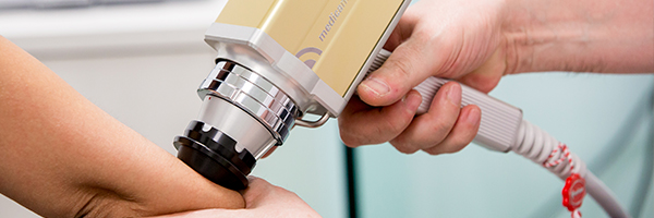
Technique used: digital dermatoscopy
Within the diagnostic procedures of skin cancer, the most important advance is the so-called digital dermoscopy, which consists of an increased vision of the person’s freckles and spots using a high resolution camera. From these increased images, we can safely and reliably diagnose whether it’s a suspicious or benign nevi and determine whether a biopsy is necessary or not.
Dr. López Gil performing a digital dermatoscopy
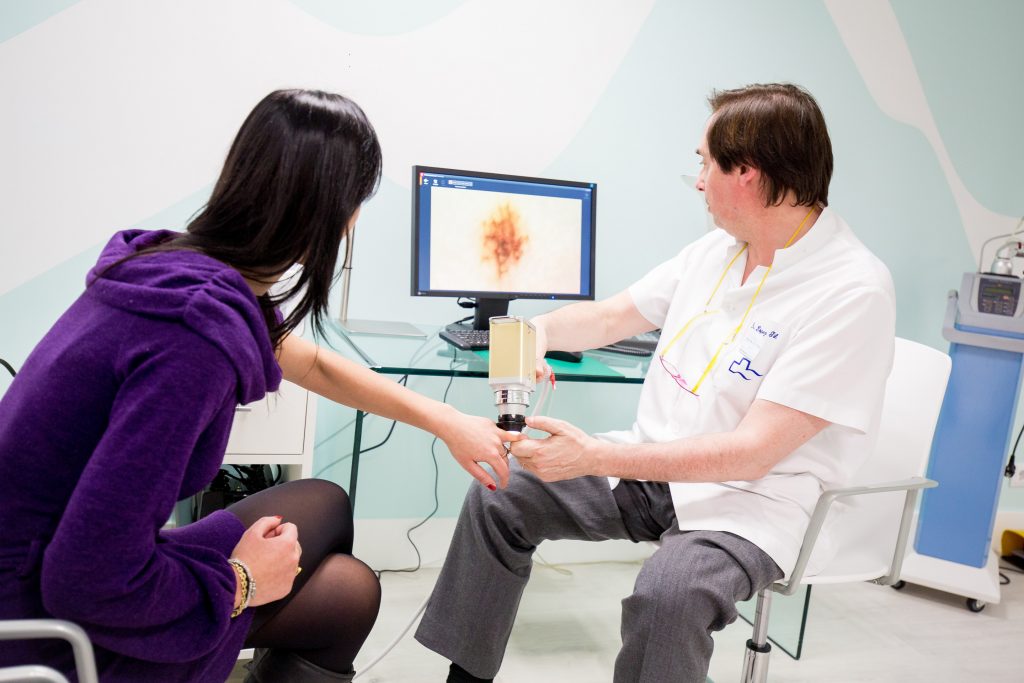
Once the digital dermatoscopy has been performed, all images taken in the patient’s history are stored in a digitized form. This photographic document allows us to compare with the maximum reliability the results obtained in the different revisions and to be able to detect in time any minimum possible change that would not have been possible to the naked eye.
Nevus change detection through digital dermatoscopy

Nevus view “ull nu”

Nevus view with digital dermoscopy
